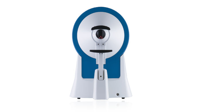
Centerfield 2
Centerfield® 2
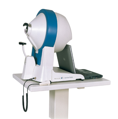
To najnowsza, zmodyfikowana wersja znanego już i uznanego polomierza projekcyjnego do badania statycznego i kinetycznego pola widzenia. Aparat umożliwia również wykonanie badań za pomocą niebieskiego bodźca na żółtym tle (test blue on yellow). Jest on w pełni zgodny z standardem Goldmanna i spełnia wymagania międzynarodowej normy perymetrycznej ISO-12866
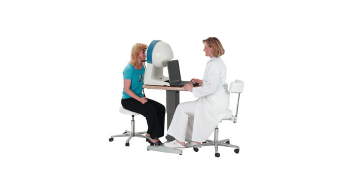
Ergonomic Design
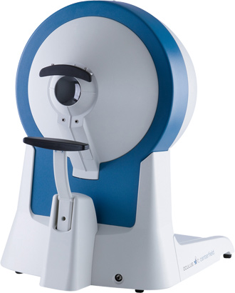
The self-contained design and light-shielded viewer allow examinations to be conducted in normally lit rooms. Its robustness and light weight make the Centerfield® 2 perimeter the ideal unit for portable use, which is often a necessity in the Occupational Health area.
Reliable Results
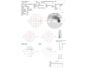
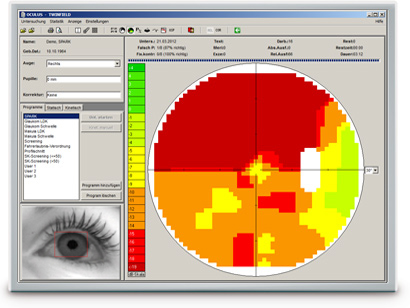
The results of the perimetric measurements are summarized in a clearly structured printout. Areas of abnormality can be quickly recognized and re-examination of these areas independent of the test point grid gives the diagnostic analysis even greater reliability.
Modern Connectivity
The Centerfield® 2 perimeter can be operated via the USB port of a notebook or PC. Together with the supplied device software, this modern solution allows you to fully utilize all of the benefits of today’s IT systems, especially the network connectivity. This guarantees both safe storage of examination data and quick access to that data when needed.
The Examination Programs
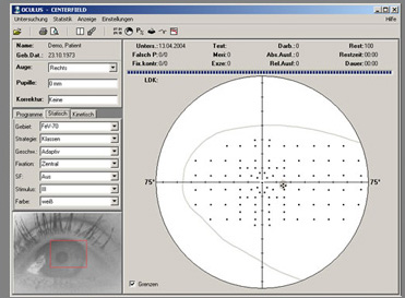
The recommended series of pre-defined test programs provided for Centerfield® 2 perimeter covers the examinations most frequently needed in practice. The “Driver’s License Regulations“ program permits occupational and company physicians to test the field of vision in accordance with applicable German regulations (FEV and amendments thereto) governing the issuance of driver’s licenses. The macula and glaucoma programs allow early detection of various retinal diseases.
The OCULUS Test Strategies
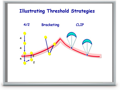
Threshold related – suprathreshold strategies have proven to be time-saving and efficient examination methods. The Centerfield® 2 perimeter uses these strategies for all screening programs, including the Driver’s License eye exams. A variety of threshold testing strategies are available for measurement of the exact numeric values of the visual thresholds.
The OCULUS Test Point Grid
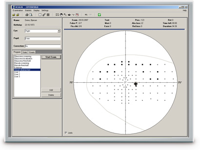
The patented projection method allows you to freely select test point grids on the Centerfield® 2. The central field of vision can be examined using functionally or orthogonally distributed test points. Grids such as the “FeV70” or “Esterman” are used for the periphery. This unlimited flexibility enables test grids to be modified if necessary.
SPARK Strategy
The SPARK examination strategy was primarily developed for glaucoma patients and is available for all OCULUS perimeters. It is based on data from more than 90 000 perimetric diagnoses and allows a very fast, yet very precise measurement of the thresholds in the central field of vision. The modular configuration of the method makes it suitable for a variety of applications:
- SPARK Precision (optional software) is the full version. The complete examination of the field of vision of glaucoma patients takes only 3 minutes per eye. The greater stability of the results allows for much better progression analysis.
- SPARK Quick is the strategy used for progression or screening examinations. The test takes just 90 seconds per eye.
- SPARK Training is ideal for patient training. The 40 seconds measurement can also be used as a screening examination.
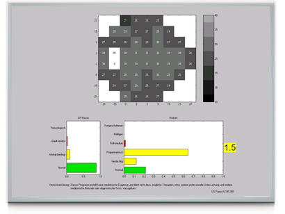
Glaucoma Staging Program (GSP)
The Glaucoma Staging Program (GSP) is useful in early detection of Glaucoma.
The GSP software grades examination findings into visual field classes (normal, glaucomatous, artifactual and neuro) based solely on their appearance. In addition, risk classes (normal, suspect, pre-perimetric, early stage, moderate and severe) are also assigned to findings of different severities that are classified as normal or glaucomatous. The examination results are presented in easy to understand green, yellow and red bar charts.
The striking novelty of the GSP is its ability to detect subtle changes in the visual field associated with early stage Glaucoma. Findings in the suspect and pre-perimetric risk classes contain reductions in the visual field that cannot be readily seen by the examiner. They usually remain undetected by the standard perimetric indices.
The Glaucoma Likelihood Index (GLI) summarizes the results of the GSP classification into a single parameter, presenting a value of between 0 (normal) and 5 (advanced Glaucoma).
The Glaucoma Staging Program (GSP) is available for the OCULUS Twinfield® 2, Centerfield® 2, Smartfield and Easyfield® perimeters. This upgrade is available for the existing units.
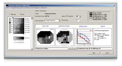
Threshold Noiseless Trend (TNT) Progression Analysis
TNT provides a quantitative, statistical analysis of the visual field examination results conducted over time. For follow-up purposes, the software uses the visual field results and patient’s threshold values supplied by 30-2, 30×24 or 24-2 grids over the entire examination period. TNT uses a specific filter to reduce the fluctuation range of the threshold values and to perform a consistent trend analysis. In conjunction with the fast SPARK strategy, the sensitivity of detecting progression of early stage glaucoma is greatly improved.
- TNT creates a concise progression analysis report containing the most important parameters (MD increase, p-values, etc.).
- TNT can differentiate between diffuse and focal progression based on the focusing index (FI) value.
- TNT applies multiple statistical criteria to establish possible progression.
- TNT displays the prognosis of the expected visual field for a patient age-group selected by the examiner.
| Programs: | Pre-defined glaucoma, macula, screening and neurological tests, user-defined tests |
| Test patterns: | Orthogonal patterns (30-2, 24-2, 30 x 24, 10-2), physiological patterns (Area 1-8), Esterman, customized patterns |
| Strategies: | Threshold strategies: OCULUS Fast Threshold, Full Threshold (4-2), CLIP; Optional: SPARK strategy, Age-adapted supra-threshold strategies: OCULUS class strategy, 2-zone, 3-zone, quantify defects |
| Examination speed: | Adaptive / fast / normal / slow / user-defined |
| Fixation control: | Through central threshold, Heijl-Krakau (using the blind spot), live video image |
| Perimeter bowl radius: | 300 mm |
| Max. eccentricity: | 36° / 70° (with fixation shift) |
| Stimulus size: | Goldmann III |
| Stimulus colour: | White, blue |
| Stimulus duration: | 200 ms / user-defined |
| Stimulus luminance range/step: | 0 – 318 cd/m² (1 000 asb) / 1 dB |
| Background luminance: | 10 cd/m² (31.4 asb) |
| Background colour: | White, yellow |
| Result display: | Greyscale, dB values (absolute/relative), symbols, probabilities, 3D plot |
| Reports: | Glaucoma Staging Program (GSP), Threshold Noiseless Trend (TNT) progression report |
| Strategies: | Automated tests along meridians with freely selectable density up to 35° |
| Stimulus speed: | 2°/s or user-defined |
| Patient positioning: | In depth adjustable head rest, in height adjustable motorized chinrest (optional) |
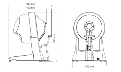
| Dimensions (W x D x H): | 398 x 503 x 580 mm (15.7 x 19.8 x 22.8 in) |
| Weight: | 12.8 kg (28.2 lbs), without chinrest 11.7 kg (25.8 lbs) |
| Power supply: | 15 V DC, 3.3 A |
| Voltage: | 100 – 240 V AC |
| Frequency: | 50 – 60 Hz |
| Recommended computer specifications: | Intel® Core™ i5, 500 GB HDD, 4 GB RAM, Windows® 7 Pro, Intel® HD Graphics 520 |
| Interface: | USB |
| Software: | Device control, patient management, backup and print software (Windows®) Built-in networking, easy EMR-integration, DICOM compatibility |

