Strona główna » Oferta » Topografia »
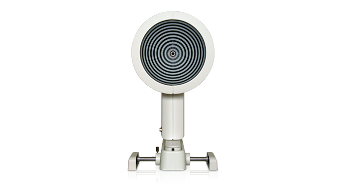
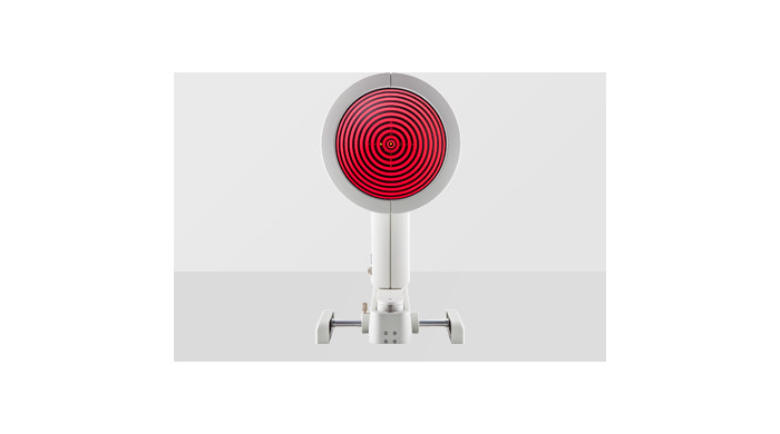
Keratograph 4
Keratograph 4
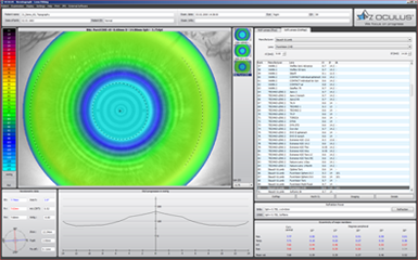
Topograf- keratograf firmy Oculus pozwala na stworzenie dokładnej mapy topograficznej i keratometrycznej przedniej powierzchni rogówki oka. Bogate oprogramowanie pozwala na analizę otrzymanego obrazu i wykorzystanie do wielu praktycznych zastosowań. Stabilna konstrukcja aparatu, prosta obsługa i niezawodne oprogramowanie pozwala na stosowanie aparatu w klinikach i szpitalach okulistycznych a także w gabinetach okulistycznych, do diagnostyki rogówki np. przy zabiegach refrakcyjnych, ocenie deformacji rogówki z analizą i oceną stożka rogówki, a także do dobierania soczewek kontaktowych (wbudowany program dobierania soczewek firmy Hecht).
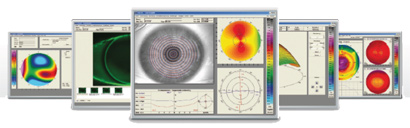
A Picture Says More Than 1 000 Words
Use the Keratograph 4 as a marketing tool and incorporate it actively into your consultations. With the Keratograph 4 software you can show images which your clients/patients have never seen before.
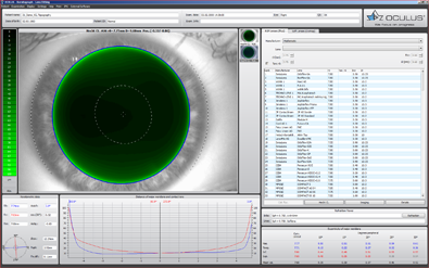
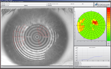
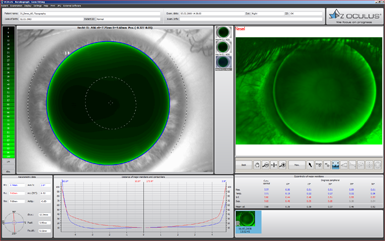
Contact Lens Fitting and Fluorescein Image Simulation
Contact lenses are on an individual basis and displayed in a list. In order to avoid taking more steps than necessary when fitting contact lenses, the fluorescein image can be simulated beforehand. The contact lens can be rotated and moved around. Fluorescein image simulation is adjusted automatically. The integrated and expandable database contains all customary types of contact lenses and is updated on a regular basis. The user can determine the order in which contact lens manufacturers appear.
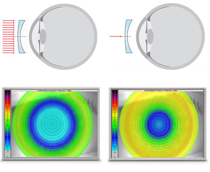
OxiMap® – Visualizing the Oxygen Transmissibility
Professional patient consultation
The cornea needs oxygen and a good oxygen supply is fundamental for the comfort of a contact lens wearer. New materialsused for soft contact lenses offer excellent oxygen transmissibility. This can be shown with the new OCULUS OxiMap® display. You can easily show these color maps to your patients and help them choose better contact lenses.
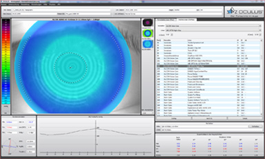
TF-Scan Makes the Tear Film Visible
This software shows the quality and quantity of the tear film.
In cases of dry eye patients and contact lens wearers, the tear film should be examined carefully. Only an intact tear film guarantees contact lens wearing comfort! The Keratograph 4 measures the tear film breakup time non-invasively (quality assessment). You can show your patient the individual tear film quality using the color maps and by taking another non-invasive measurement to determine the amount of tear film (tear film quantity).
TF-Scan Makes the Tear Film Visible
Patient consultation made easy. This software shows the quality and quantity of the tear film.
In cases of dry eye patients and contact lens wearers, the tear film should be examined carefully. Only an intact tear film guarantees contact lens wearing comfort! The Keratograph 4 measures the tear film breakup time non-invasively (quality assessment). You can show your patient the individual tear film quality using the color maps. In addition, you can take another non-invasive measurement to determine the amount of tear film (tear film quantity).

Tear Film Quality (NIKBUT)
The OCULUS Keratograph 4 determines the break-up time using the NIKBUT procedure (non-invasive-Keratograph-break-up time).
Different Ways of Determining Pupil Reaction:
- Examination of the pupil reaction both with and without glare
- Examination using two different glare stimulus powers
- Clear presentation of the results in graphic form: pupil changes over a period of time; minimum, maximum and mean pupil diameter, incl. standard deviation
- Comparison views possible
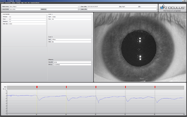
Pupillometry
Using the “Pupillometry” option, the reaction of the pupil, can be checked with and without blinding. This builds the basis for selecting the proper treatment zone for laser controlled surface ablation, multifocal contact lenses or premium IOLs. The pupil reaction of the two eyes can be compared.
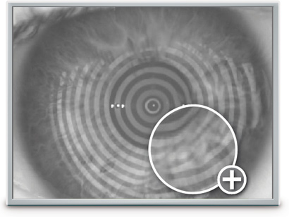
Optional Imaging-Software
The Imaging-Software takes real-time fluo-images or fluo-clips of the contact lens. The high resolution clip shows the movements of the contact lens on the cornea. A side-by-side comparison displays the simulated fluo image of the software and the real time fluo image and the clip.
Essential for:
- Demonstration of the fit and mobility of contact lenses
- Assessment of the static fluo-image
- Assessment of the fit of the contact lenses with different pupil diameters
- Side-by-side comparison of fluo-image simulations to real-time fluo-images
- Choice of the final contact lense
- Expertise and customer long term relationship
OCULUS OxiMap®
The OxiMap® presents a color map of the oxygen transmissibility of soft contact lenses based on the lens power, which is easy to understand – even for your customers!
How Much Oxygen Really Reaches the Cornea?
Until now, only the oxygen transmissibility values for the center of a contact lens with -3.0 D were available. The OxiMap® shows the oxygen transmissibility depending on the lens material and the lens thickness. The OxiMap® is available for the most frequently sold spherical soft contact lenses. This impressive tool assists you in helping your patients select the most suitable contact lens.
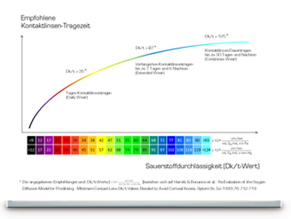
Plain and Comprehensive Visualization Assures Patient Loyalty!
Contact lenses act as a potential barrier to oxygen transport even when the eyes are open to the atmosphere. Long hours of wearing comfort can only be guaranteed with a sufficient oxygen supply. The color representation of the various terms of oxygen transmissibility is based on international recommendations for daily, extended and continuous wear.
Compare Different Soft Contact Lenses!
The OxiMap® is individually adapted based on the lens power and supports you in your consultation with the patient and helps you to choose the most suitable contact lens. New materials used for soft contact lenses provide excellent oxygen transmissibility.
OxiMap® was developed in close cooperation with JENVIS Research and the University of Applied Sciences in Jena.
| Measurement range | 3 – 38 mm (0.1 – 1.5 in), 9 – 99 D |
| Precision | ± 0.1 D |
| Reproducibility | ± 0.1 D |
| Number of rings | 22 |
| Working distance | 80 mm |
| Number of measuring points | 22 000 |
| Camera | digital CCD camera |
| Source of illumination | placido illumination: red 650 nm imaging illumination: blue 465 nm (UV-free) pupillometer illumination: infrared 880 nm |
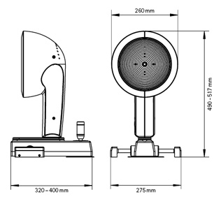
| Dimensions (W x D x H) | 275 x 320 – 400 x 490 – 517 mm |
| Weight (Measuring head) | 2.3 kg (5.1 lbs) |
| Weight (with base) | 5.3 kg (11.7 lbs) |
| Power Consumption max. | 30 W |
| Voltage | 100 – 240 V AC |
| Frequency | 50 – 60 Hz |
| Minimum PC requirements | Intel® Core™ i5, 1 TB HDD, 8 GB RAM, Windows® 7 – Windows® 10 |
| Recommended screen resolution | 1920 x 1200 pixel |