Strona główna » Oferta » Pentacam »
Pentacam
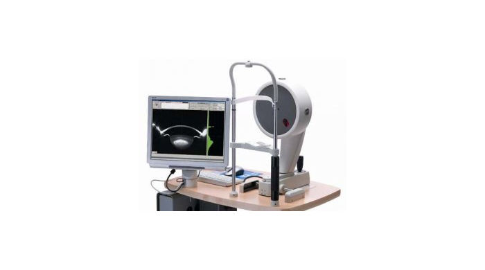
Pentacam®
Pentacam to obrotowa kamera Scheimpfluga, która uzyskuje obrazy Scheimpfluga przedniego odcinka oka.Technika Scheimpfluga zapewnia ostre i wyraziste obrazy, które dostarczają informacji o strukturach od przedniej powierzchni rogówki do tylnej torebki soczewki. Najważniejszymi zaletami procesu obrazowania rotacyjnego jest możliwość precyzyjnego pomiaru środkowej części rogówki, korekcja względem ruchów oka, łatwa dla pacjenta fiksacja i bardzo krótki czas badania. Pentacam tworzy trójwymiarowy model matematyczny przedniego odcinka, dostarczając następujących danych:
- mapy topograficzne i elewacyjne przedniej i tylnej powierzchni rogówki
- pachymetria rogówkowa od rąbka do rąbka
- trójwymiarowa analiza komory (mapa ACD, kąt komory, objętość komory itp.)
- gęstość soczewki (ilościowa ocena przepuszczalności świetlnej krystalicznej części rogówki i soczewek IOL)
- tomografia
- dokładniejsze obliczenia IOL dla pacjentów po zabiegach LASIK, PRK i RK.
Podsumowując wszystkie jego cechy, aparat Pentacam jest najlepszym urządzeniem diagnostycznym dla następujących zastosowań:
- Chirurgii refrakcyjnej rogówki,
- Chirurgii zaćmy,
- przesiewowych badań pod kątem jaskry,
- ogólnych badań przesiewowych

A complete diagnosis in just the bat of an eye.
Within just two seconds the Pentacam® supplies you with precise diagnostic data on the entire anterior eye segment. The degree of corneal or crystalline lens density is made visible by the light scattering properties of the crystalline lens and is automatically quantified by the software. Measurement of the anterior and posterior corneal surfaces supplies the total refractive power as well as the thickness of the cornea over its entire area. The data on the posterior surface provide optimal assistance in the early detection of ectatic changes.
The rotating scan supplies a large number of data points in the center of the cornea. A supplementary pupil camera captures eye movements during the examination for subsequent automatic correction of measured data.
Versatile Basic Software
Even the basic software offers a vast range of functions.
- Fast Screening Report
- Contact Lens Fitting
- Belin/Ambrósio Enhanced Ectasia Display
- Topographical Keratoconus Classification (TKC)
- Tomographical Keratoconus Classification
- Belin ABCD Keratoconus Staging
- Belin ABCD Progression Display
- Qualitative assessment of the cornea
- Topography and Elevation Maps of the Anterior and Posterior Corneal Surface
- overall pachymetry
- Glaucoma screening:
- pachymetry-based IOP correction
- chamber angle and chamber volume
- Elevation data
- Comparative displays for follow-up
- Comparison and superimposition of Scheimpflug images
Pełną specyfikację produktu znajdą Państwo pod tym adresem.
General Overview
Uses
- Comprehensive overview
- Customer consultation
Details
The General Overview provides important information on the keratometry and pachymetry of the cornea in concise numerical form. The Scheimpflug image provides opticians/optometrists and their customers with intuitive representations of opacities of the cornea or lens (cataract) or of the position of existing IOLs. The anterior chamber is described in terms of anterior chamber depth, volume and angle. When combined with IOP tonometry readings corrected for corneal thickness the General Overview permits an assessment of glaucoma risk. It also allows the display of all colour maps.