Smartfield
Smartfield
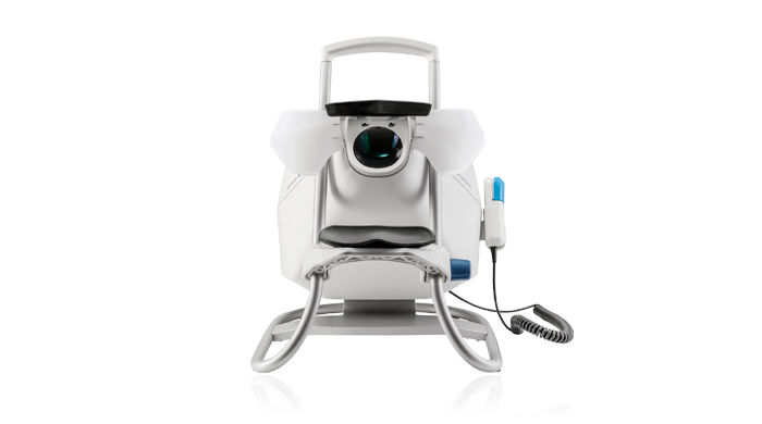
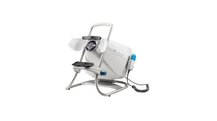
The OCULUS Smartfield is a compact visual field device purposefully optimized for monitoring functional impairment in glaucoma. Based on an ultra-high-luminance LCD screen it performs standard automated perimetry of the central visual field and beyond. Despite its small size, the Smartfield perimeter offers a comprehensive clinical solution in visual field testing for any glaucoma practice.
Ergonomic Design
The OCULUS Smartfield stands out with a very small footprint, which is smaller still than that of the Easyfield®. Its closed design and light-protected viewer permit examinations in rooms with normal lighting conditions. Its low weight and the practical carrying handle make the Smartfield perimeter convenient for portable use. The height-adjustable double chin rest and measuring head allow for better positioning of the patient and improved comfort. The absence of moving parts guarantees a long product lifetime.

Easy Operation
The Smartfield perimeter is designed for operation via an external computer (notebook or PC) connected to the common office data network. This ensures network availability of examination data. The translucent lateral eye shields render the use of an eye patch during the examination unnecessary, thus saving valuable time in preparation for the test.
Increased Diagnostic Reliability
Powerful test strategies such as SPARK and other assessment tools contribute to the great diagnostic utility of the Smartfield perimeter. The enhanced Glaucoma Staging System (GSS 2) of Dr. Brusini and the Glaucoma Staging Program (GSP) offer extended support in single field analysis. Structure-function relationships are predicted by the new PATH evaluation module. The Threshold Noiseless Trend (TNT) module performs efficient progression analysis. Examination quality is ensured by a patented fixation control algorithm, a high resolution video camera for eye monitoring and the various built-in catch trials.
Examination Programs
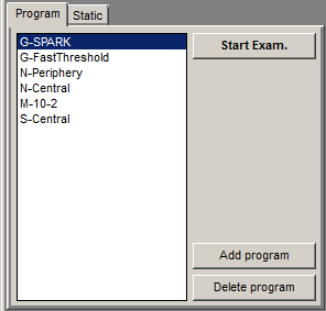
The Smartfield is equipped with a set of predefined programs for the most frequently needed examination routines of the central visual field or the macular area. A default program for the periphery is also included. The program list can be easily extended according to specific requirements by combining the available test patterns and test strategies.
OCULUS Test Strategies
The Smartfield features the innovative SPARK test strategy for faster and more stable threshold measurements in glaucoma patients. Rounding off the testing capabilities of the Smartfield is the complete set of traditional OCULUS test methods, including threshold and suprathreshold strategies. The classical 4-2 dB staircase strategy (Threshold) uses two reversals in the patient’s answer to deliver a threshold value in each tested point. The OCULUS Fast Threshold strategy is a clever improvement over the classical procedure which reduces examination time by using variable steps and taking advantage of already measured locations. Suprathreshold strategies like the 2-zone or 3-zone strategy offer a quick overview of the visual field.

OCULUS Test Patterns
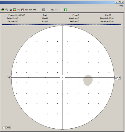
The grid corresponding to the SPARK examination strategy (30×24) is the preferred pattern in the Smartfield. However, other commonly used patterns such as 24-2 or 10-2 are readily available. Custom patterns for the central visual field can be easily created. The modular structure of the test programs allows for all patterns to be examined using any available standard strategy.
Results Printout
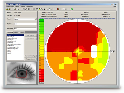
Measurement results of the Smartfield perimeter are commonly summarized in a standard printout. For suprathreshold tests a qualitative overview is printed, whereas for threshold examinations all clinically relevant data are shown in various representations. Besides the standard printout the Smartfield software features various assessment displays.
SPARK Strategy
The SPARK examination strategy was primarily developed for glaucoma patients and is available for all OCULUS perimeters. It is based on data from more than 90 000 perimetric diagnoses and allows a very fast, yet very precise measurement of the thresholds in the central field of vision. The modular configuration of the method makes it suitable for a variety of applications:
- SPARK Precision (optional software) is the full version. The complete examination of the field of vision of glaucoma patients takes only 3 minutes per eye. The greater stability of the results allows for much better progression analysis.
- SPARK Quick is the strategy used for progression or screening examinations. The test takes just 90 seconds per eye.
- SPARK Training is ideal for patient training. The 40 seconds measurement can also be used as a screening examination.
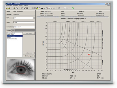
Glaucoma Staging System (GSS 2) per Brusini
The Glaucoma Staging System (GSS 2) per Brusini classifies visual field defects based on the perimetric indices MD (Mean Deviation) and PSD (Pattern Standard Deviation). The representative point of the examination is placed in a chart in accordance with the values of these indices. The chart shows defined areas for the different stages of disease (Stage 0 – Stage 5). It also separates generalized, localized and mixed defects.
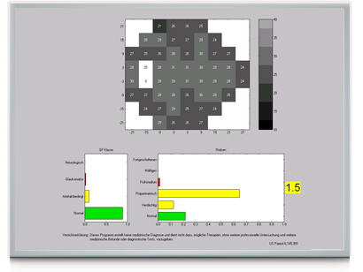
Glaucoma Staging Program (GSP)
The Glaucoma Staging Program (GSP) is useful in early detection of Glaucoma.
The GSP software grades examination findings into visual field classes (normal, glaucomatous, artifactual and neuro) based solely on their appearance. In addition, risk classes (normal, suspect, pre-perimetric, early stage, moderate and severe) are also assigned to findings of different severities that are classified as normal or glaucomatous. The examination results are presented in easy to understand green, yellow and red bar charts.
The striking novelty of the GSP is its ability to detect subtle changes in the visual field associated with early stage Glaucoma. Findings in the suspect and pre-perimetric risk classes contain reductions in the visual field that cannot be readily seen by the examiner. They usually remain undetected by the standard perimetric indices.
The Glaucoma Likelihood Index (GLI) summarizes the results of the GSP classification into a single parameter, presenting a value of between 0 (normal) and 5 (advanced Glaucoma).
The Glaucoma Staging Program (GSP) is available for the OCULUS Twinfield® 2, Centerfield® 2, Smartfield and Easyfield® perimeters. This upgrade is available for the existing units.
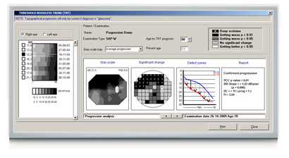
Threshold Noiseless Trend (TNT) Progression Analysis
TNT provides a quantitative, statistical analysis of the visual field examination results conducted over time. For follow-up purposes, the software uses the visual field results and patient’s threshold values supplied by 30-2, 30×24 or 24-2 grids over the entire examination period. TNT uses a specific filter to reduce the fluctuation range of the threshold values and to perform a consistent trend analysis. In conjunction with the fast SPARK strategy, the sensitivity of detecting progression of early stage glaucoma is greatly improved.
- TNT creates a concise progression analysis report containing the most important parameters (MD increase, p-values, etc.).
- TNT can differentiate between diffuse and focal progression based on the focusing index (FI) value.
- TNT applies multiple statistical criteria to establish possible progression.
- TNT displays the prognosis of the expected visual field for a patient age-group selected by the examiner.
Predicting Anatomy from Thresholds (PATH)
Functionality of a retinal location obviously depends on the underlying anatomical structure of the retina in that location. One characteristic feature of glaucoma is that there is a close connection between sensitivity levels of given retinal locations (as described by visual field measurements) and anatomical structures related to the optic nerve head. The merit of PATH is that it provides an estimate of the retinal nerve fibre layer (RNFL) thickness around the optic nerve head and the relative area of the neuroretinal rim based solely on the results of visual field measurements performed with the SPARK examination strategy. This novel approach of predicting structure from function is made possible by the high reproducibility of the SPARK visual field exams.
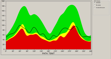
Predicting RNFL thickness
Working from the results of SPARK perimetry, RNFL thickness is predicted at 25 points of the TSNIT (Temporal – Superior – Nasal – Inferior – Temporal) circle around the optic nerve head. The value at each point is calculated from functional data selected for relevance. This selection is based on an objectively automated machine learning algorithm and does not rely on other findings such as on the correspondence between nerve fibre pathways and visual field areas.
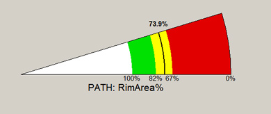
Predicting the relative area of the neuroretinal rim
A linear combination of relevant threshold results is used in order to predict the ratio between the neuroretinal rim area and the total area of the optic disc. The result is compared to normative data and expressed as a percentage of the population average value (normalized to 100%).
| Programs | Pre-defined glaucoma, macula, screening and neurological tests user-defined tests |
| Test patterns | 30×24 (SPARK), 24-2, 10-2, customized patterns |
| Strategies | Threshold strategies: SPARK Precision, SPARK Quick, OCULUS Fast Threshold, Full Threshold (4/2), Age-adapted suprathreshold screening (2-zone, 3-zone) |
| Examination speed | Adaptive/fast/normal/slow/user-defined |
| Fixation control | Through central threshold, Heijl-Krakau (using the blind spot), live video image |
| Result display | Greyscale, dB values (absolute/relative), symbols, probabilities, 3D plot |
| Reports | Enhanced Glaucoma Staging System (GSS 2), Glaucoma Staging Program (GSP), PATH function-structure analysis, Threshold Noiseless Trend (TNT) progression report |
| Stimulus viewing distance | Infinity |
| Max. eccentricity horizontal/vertical | 30°/25° (60°/50° with fixation shift) |
| Stimulus size | Goldmann III |
| Stimulus colour | White |
| Stimulus duration | 200 ms/user-defined |
| Threshold range/step | 0.8 – 3 180 cd/m2 (2.5 – 10 000 asb), 0 – 36 dB/1 dB |
| Background luminance | 10 cd/m2 (31.4 asb) |
| Patient positioning | Height-adjustable measuring head, adjustable chin rest, double head rest |
| Software | Device control, patient management, backup and print software (Windows®) Built-in networking, easy EMR-integration, DICOM compatibility |
| Dimensions (W x D x H) | 332 x 418 to 477 x 402 mm (13.1 x 16.5 to 18.8 x 15.9 in) |
| Weight | 7.6 kg (16.8 lbs) |
| Max. power consumption | 30 W |
| Voltage | 100 – 240 V AC |
| Frequency | 50 – 60 Hz |
| Recommended computer specifications | Intel® CoreTM i5, 500 GB HDD, 4 GB RAM, Windows® 7 Pro, Intel® HD Graphics 520 |
