Strona główna » Oferta » Topografia »
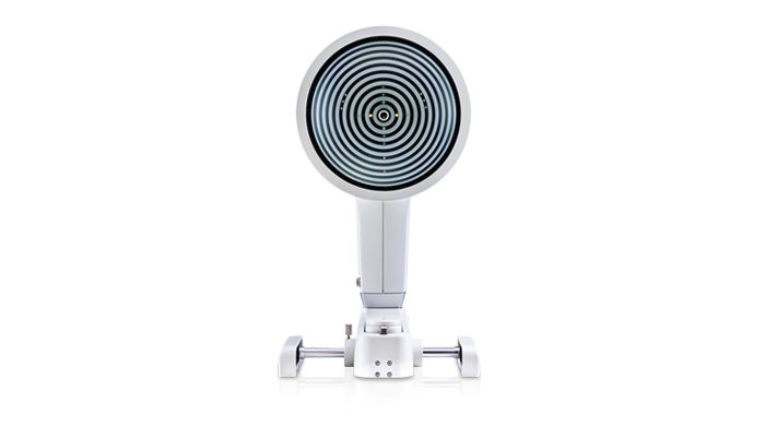
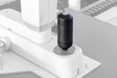
Keratograph 5M
Keratograph 5M
The OCULUS Keratograph 5M is an advanced corneal topographer with a built-in real keratometer and a color camera optimized for external imaging. Unique features include examining the meibomian glands, non-invasive tear film break-up time and the tear meniscus height measurement and evaluating the lipid layer.
Gain Trust
With the Keratograph 5M, you can show your patients images they have never seen before. Gain patient trust by providing professional consultation during examinations and follow-ups.
Easy-to-Understand Presentation
Actively integrate the Keratograph into your consultation. Many easy to understand displays support you in communication and patient education. Use your Keratograph 5M as a marketing tool to make your services transparent!
New Joystick for Keratograph 5M: Now with Release Function
The new joystick for the Keratograph 5M allows you to capture images and video sequences directly from the device. No need to use the mouse: your hand just stays on the joystick when you capture images or videos.
The new joystick communicates via a smart Bluetooth interface. And it does away with the need for cumbersome power cabling. The joystick switches to sleep mode function when not in use to extend battery life.
Switching to the new joystick is quick and simple. Watch the video below to see how easy it is!
Installing and using the new joystick
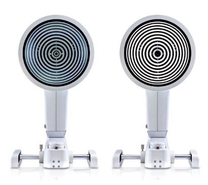
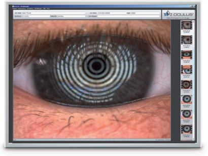
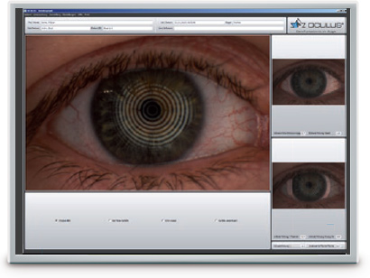
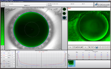
Measurements with Placido Ring Illumination
Thousands of measuring points are used to measure the whole surface of the cornea. A white ring illumination is used for this purpose. An infrared ring illumination is also provided for analysis of the tear film to prevent glare-related reflex secretion.
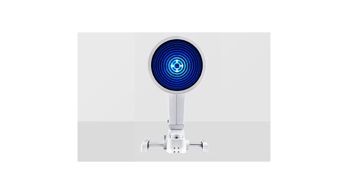
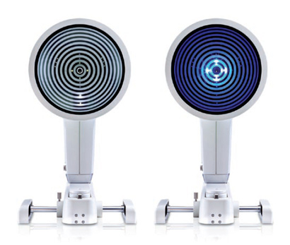
Measurements with Light Emitting Diodes
The perfect illumination has been integrated for every function of the Keratograph 5M: White diodes for the tear film dynamics, blue diodes for fluo-images, infrared diodes for Meibography.
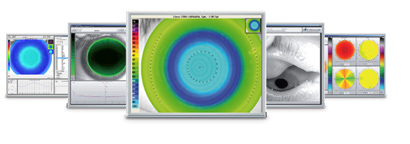
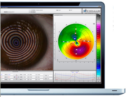
JENVIS Pro Dry Eye Report: Your Professional Dry Eye Assistant
Find the cause of dry eye syndrome quickly and reliably. The new JENVIS Pro Dry Eye Report in the Keratograph 5M will help you as you go along. Perform a comprehensive screening, using the measuring results as a basis for diagnosing dry eye syndrome. All results are documented in accordance with the Medical Products Law and summarized for your patient in a neat and easily understandable printout.
JENVIS Pro Dry Eye Software – The Dry Eye Analysis Tool
Make use of all the advantages of the new JENVIS Pro Dry Eye Report in the Keratograph 5M: efficient screening, well-founded measurement results and greater patient loyalty.
Screening for dry eye syndrome should be a routine part of every refraction.
TF-Scan
The tear film is assessed using either white or infrared illumination. The new high-resolution color camera makes even the finest of structures visible. In addition to NIKBUT (Non-Invasive Keratograph Break-Up Time) and measurement of the meniscus tear, the TF-Scan can also make an assessment of the lipid layer and the tear film dynamics. Tear film analysis with the OCULUS Keratograph 5M is non-invasive and is conducted without any additional tools.
NIKBUT
Assessment of the Tear Film Break-Up Time
The tear film break-up time is measured non-invasively and fully automatically. The new infrared illumination is not visible to the human eye. This prevents glare during the examination. The TF-scan presents the results in a way that is easy for you and your customers to understand.
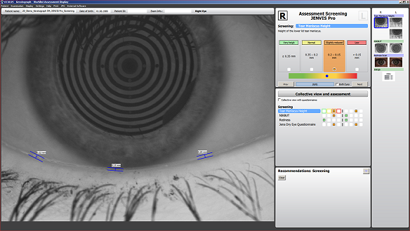
Meniscus Tear Height
Assessment of the Tear Film Quantity
The height of the torn meniscus can be precisely measured with an integrated ruler and various magnification options, and its development along the edge of the bottom lid can be assessed. The results are saved to the patient file.
Lipid Layer
Assessment of the Interference Phenomenon
The interference colors of the lipid layer and their structure are made visible and can be recorded. The thickness of the lipid layer is assessed based on the structure and color.
TF Dynamics
Assessment of the Particle Flow
The video recording, with up to 32 frames per second, enables the observation of the tear film particle flow, from which conclusions regarding the viscosity of the tear film can be drawn.
R-Scan
Automatic Classification of Redness
Automatic classification of the bulbar redness. Conjunctival redness used to be assessed subjectively and depended on the examiner. The R-Scan is the first module that automatically and objectively documents and classifies the bulbar and limbal degree of redness. The R-Scan detects the blood vessels in the conjunctiva and evaluates the degree of redness. This automatic assessment spares you the time and effort of having to make manual comparisons using classification sheets.
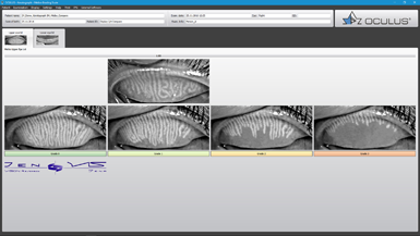
Meibo-Scan
Meibography of the Top and Bottom Eyelid
The multi-functionality of the Keratograph 5M easily and efficiently integrates difficult examinations such as meibography. The dysfunction of meibomian glands is the most frequent cause of dry eye. Morphological changes in the gland tissue are made visible using the Meibo-Scan and can be classified with the JENVIS Meibo Grading Scales.
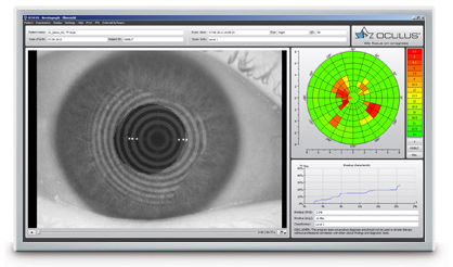
Pupillometry
Indispensable for:
- Fitting multi-focal contact lenses
- Exact determination of the treatment area for refractive surgery
- Seamless integration into the existing Keratograph software
- The infrared camera installed in the Keratograph delivers images of the patient’s pupil, which are used as the basis for the measurements
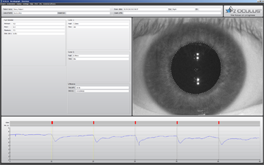
Different Ways of Determining Pupil Reaction:
- Examination of the pupil reaction both with and without glare
- Examination using two different glare stimulus powers
- Clear presentation of the results in graphic form: pupil changes over a period of time; minimum, maximum and mean pupil diameter, incl. standard deviation
- Comparison views possible
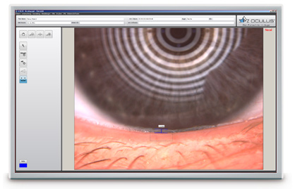
Imaging-Software
The Imaging Software is used to record video and image files. In addition to viewing the videos and single images by themselves, you can also compare the recordings with the simulated fluorescein-images of the measured eye.
How Does That Work?
- Simply integrate the Imaging Software into the Keratograph software
- Blue light-emitting diodes in the illumination beam path stimulate the fluorescein
- Yellow filters are integrated into the observation beam path
Result: Static fluorescein-images and videos can be recorded under slit lamp conditions!
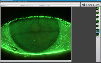
Indispensable for:
- Demonstrating the fit of contact lenses
- Assessment of the static fluo-image
- Assessment of the fit of contact lenses at different pupil sizes
- Comparison of fluo-image simulations with real-time fluo-images
- Selection of the best possible contact lens
- Consultation and customer retention
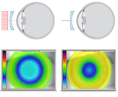
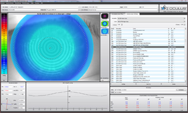
OCULUS OxiMap®
The OxiMap® presents a color map of the oxygen transmissibility of soft contact lenses based on the lens power, which is easy to understand – even for your customers!
Oxygen Transmissibility of Soft Contact Lenses Made Visible
An intact tear film and a good supply of oxygen to the cornea are an absolute necessity for wearing contact lenses comfortably. The OxiMap® presents a color map of the oxygen transmissibility of soft contact lenses based on the lens power – which even your customers will understand!
Easy-to-Understand Patient Education
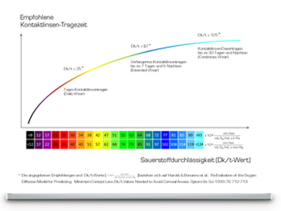
The oxygen transmissibility (Dk/t) depends on the material the contact lens is made of and on its thickness. In the past, only the manufacturers’ specifications about the oxygen transmissibility in the center of a -3,00 dpt contact lens were available. With the OxiMap®, you now get a graphic visualization of the Dk/t values over the whole area, based on the lens thickness.
The Dk/t values are color-coded, whereby black represents an oxygen supply lower than that present in a closed eye. To preserve the integrity of the cornea when wearing contact lenses, minimum Dk/t values are recommended based on the length of time the contact lenses are to be worn. The OxiMap® color-coding is based on these international recommendations.
Compare Different Soft Contact Lenses!
The OxiMap® is individually adapted based on the lens power and supports you in your consultation with the patient and helps you to choose the most suitable contact lens. New materials used for soft contact lenses provide excellent oxygen transmissibility.
OxiMap® was developed in close cooperation with JENVIS Research and the University of Applied Sciences in Jena.
| Accuracy: | ± 0.1 D |
| Reproducibility | ± 0.1 D |
| Number of rings: | 22 |
| Working distance: | 78 – 100 mm |
| Number of evaluated data points | 22 000 |
| Camera: | Digital CCD camera |
| Light source: | Placido illumination: White Placido illumination: Infrared 880 nm Fluorescein illumination: Blue 465 nm Meibography: Infrared 840 nm Tear film dynamics: White Pupillometer illumination: Infrared 880 nm |
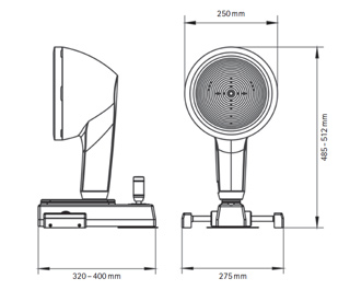
| Dimensions (W x D x H): | 275 x 320 – 400 x 485 – 512 mm |
| Weight | Measuring head: 3.2 kg (7.1 lbs) With base: 6.1 kg (13.5 lbs) |
| Max. power consumption | 18 W |
| Voltage | 90 – 264 V AC |
| Frequency | 47 – 63 Hz |
| Recommended computer specifications | CPU Intel® Core i5-7600, 8 GB RAM, 1 TB, Windows® 10 Pro |
| Recommended screen resolution | 1920 x 1200 pixel |