Strona główna » Oferta »
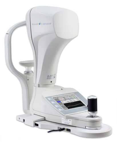
Corvis ST
Corvis® ST
Bezkontaktowy tonometr z oceną i wizualizacją własności biomechanicznych rogówki w technologii oprtej na zasadzie kamery Scheimpfluga.
Zastosowanie aparatu Corvis® ST
Określanie rzeczywistego ciśnienia wewnątrzgałkowego IOP
- Corvis® ST mierzy IOP, grubość i właściwości biomechaniczne rogówki.Opierając się na tych danych aparat podaje rzeczywistą wartość IOP.
- Corvis® ST określa wpływ właściwości biomechanicznych na standardowy pomiar IOP.
Wykrywanie ektazji rogówki
- Zarówno sztywność jak i lepkość rogówki są redukowane przez ekstazję.
- Oba czynniki mogą być analizowane jednocześnie przy użyciu Corvis® ST.


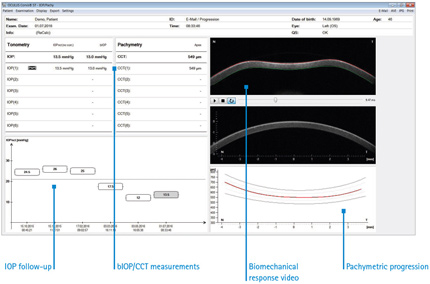
Biomechanical corrected IOP (bIOP)
More accurate IOP readings, less dependent on biomechanical properties and corneal thickness. The data are easy to read and to interpret, also the IOP follow-up is neatly arranged. IOP correction is based on corneal thickness, age and the biomechanical response of the cornea. Due to the measurement principle, the IOP measurements are not influenced by tear film.
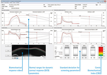
Corvis Biomechanical Index (CBI)
Comprehensive biomechanical screening and keratoconus detection. The Vinciguerra Screening Report displays the patient’s results in comparison with normative values, presented in easy-to-grasp charts.
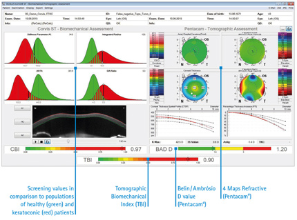
Tomographic Biomechanical Index (TBI)
Integration of Pentacam® data for a combined tomographic and biomechanical analysis. The best of two worlds: TBI is calculated using an artificial intelligence approach to optimize ectasia detection. By combining tomographic data from the Pentacam® with biomechanical data from the Corvis® ST one can further improve sensitivity and specificity in the detection of patients with a significant risk for developing ectasia after refractive surgery.
Corneal Visualization
Scheimpflug Technology
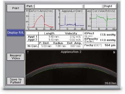
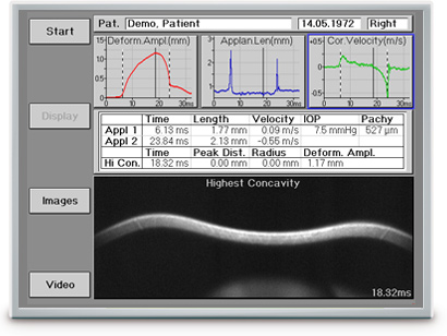
The revolutionary Corvis® ST records the reaction of the cornea to a defined air pulse using a newly developed high-speed Scheimpflug camera. This camera captures over 4 300 images per second, permitting highly precise measurement of IOP and corneal thickness. Based on a video of 140 images, taken within 31 ms after onset of the air pulse, the Corvis® ST provides a detailed assessment of corneal biomechanical properties.
The information obtained on the biomechanical response of the cornea is used to calculate a biomechanically corrected IOP (bIOP). Furthermore it allows ectatic diseases such as keratoconus to be detected at a very early stage. Biomechanical properties also play an important role in the development and progression of glaucoma.
Corvis® ST – the tool of tomorrow today
The Corvis® ST is now available in various software versions. You can start making economic use of it now. You will receive a combined Tonometer and Pachymeter complete with video function and a new email function designed for sending videos to patients´ mobiles. You can upgrade later on, adding information on biomechanical properties of the cornea from the Dynamic Corneal Response Display.
| Applications | Corvis® ST |
| IOD/Pachymetry | |
| Pachymetric Progression | |
| High-speed video of the cornea | |
| Email function for sending high-speed video | |
| DCR-Software incl. CBI and TBI | optional |
DCR: Dynamic Corneal Response
The DCR Display gives you various insights into the biomechanical properties of the cornea. The software provides detailed analysis of the Scheimpflug images taken during deformation of the cornea. The parameters which describe the deformation characteristics give information about corneal biomechanics. Also included are the Corvis Biomechanical Index (CBI) and Tomographic Biomechanical Index (TBI).
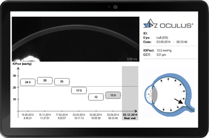
Emailing videos to patients’ mobiles
This new function allows you to email values for intraocular pressure and corneal thickness as well as impressive videos to your patients. Videos include a feature for reminding patients of upcoming follow-up exams. This way you can convince them – in particular the young ones – of the necessity for glaucoma screenings and regular follow-up exams. The high-end diagnostics provided by the Corvis® ST will put the focus on your performance and high-tech equipment.
Measurement and Display Options
- IOP measurement
- Measurement of the corneal thickness
- Scheimpflug images of the 1st and 2nd applanation of the cornea
- Slow-motion video of the corneal deformation as a result of the air pulse
- Non-contact tonometer in combination with an ultra-high-speed camera for visualization of the deformation of the cornea in reaction to an air pulse (4 330 frames / sec.)
Why are biomechanical properties so important?
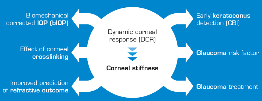
The role of the Corvis® ST in glaucoma diagnostics
The Corvis® ST measures IOP, corneal thickness and the deformation response of the cornea. Based on these data it has the potential to determine an IOP value that is uninfluenced by corneal biomechanical properties.
Screening for corneal ectasia
Corneal ectasia lead to changes in the viscoelastic properties of the cornea. These changes can be analyzed based on deviations in deformation response parameters relative to normal eyes.
Visualizing the effect of corneal crosslinking
Changes in deformation characteristics due to corneal crosslinking can also be measured with the Corvis® ST. The Corvis® ST has the potential to quantify the effect of crosslinking on the biomechanical properties of the cornea.
| Measurement range | 6 – 60 mmHg |
| Measurement distance | 11 mm (0.4 in) |
| Inner Fixation Light | Red LED |
| Frame rate | 4 330 images per sec |
| Measurement range | 8.5 mm (0.3 in) horizontal coverage |
| Pachymeter measurement range | 200 – 1 200 μm |
| Measuring points | 576 per image (80 640 per examination) |
| Source of light | Blue LED (470 nm UV free) |
| Voltage | 100 – 240 V AC |
| Frequency | 50 – 60 Hz |
| Max. power consumption | 26 W |
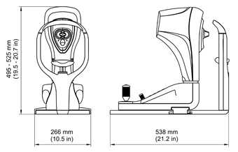
| Dimensions (W x D x H) | 266 x 538 x 495 – 525 mm (10.5 x 21.2 x 19.5 to 20.7 in) |
| Weight | 14 kg (30.8 lbs) |